

Spinal surgery is a major branch of neurosurgery and orthopedics,
primarily addressing conditions such as spinal degenerative diseases, spinal trauma, tumors, infections, and spinal deformities through surgical intervention. With the advancement of precision medicine and minimally invasive spinal surgery, an increasing number of cervical, thoracic, and lumbar procedures are being performed using microsurgical techniques to achieve smaller incisions, more precise decompression, and faster postoperative recovery.
Including these spinal surgeries
Anterior Cervical Discectomy and Fusion (ACDF), Lumbar Microdiscectomy, Laminectomy, Transforaminal / Posterior Lumbar Interbody Fusion (TLIF/PLIF), Intraspinal Tumor Resection, and other complex spinal surgeries.
These operations often require working within a narrow and deep surgical field. Surgeons must accurately identify crucial structures such as the dural sac, spinal cord, nerve roots, intervertebral discs, and paravertebral blood vessels, ensuring adequate decompression while maximizing the protection of neural tissue function. Consequently, high-quality surgical visualization has become a fundamental element for enhancing surgical precision and safety.
Our Technology's ANIM-Q Series Surgical Microscope integrates 4K ultra-high-definition 3D imaging, an extra-long working distance, digital exoscopy, and intelligent image management. It provides a more open, efficient, and precise digital surgical visualization solution for modern spinal surgery, helping surgeons meet the challenges of complex spinal microsurgical procedures.
4K ultra-high-definition 3D imaging
extra-long working distance

Key challenges in spinal surgery include:
Maintaining a clear and stable microscopic view within a deep and narrow surgical field Accurately distinguishing the spinal cord, dural sac, nerve roots, and surrounding soft tissue structures Preserving excellent three-dimensional spatial perception and depth judgment under complex anatomical conditions Providing ample working space for long instruments to enhance maneuverability in deep surgeries Improving surgeon ergonomics during prolonged microscopic procedures to reduce neck and shoulder fatigue Meeting the needs for team collaboration, intraoperative teaching, and digital image documentation in complex procedures
Digital Surgical Vision Platform for Precision Spinal Microsurgery
Addressing the clinical demands of spinal microsurgery—such as "deep surgical fields, delicate manipulation, neural protection, and team collaboration"—Our Technology introduces the ANIM-Q Series Surgical Microscope. By integrating digital visualization technologies with ergonomic design, it offers a comprehensive solution for spinal surgery covering observation, manipulation, teaching, and image management.

Dual-Channel 4K 3D Imaging Technology
The ANIM Series employs dual-channel 4K ultra-high-definition 3D imaging technology, simultaneously capturing left and right parallax information to provide the surgeon with a true, natural three-dimensional surgical field. 4K 3D imaging more accurately represents the deep tissue layers of the spine, enhancing spatial identification of critical anatomical structures like nerve roots, the dural sac, and intervertebral discs. This facilitates more precise decompression, dissection, and microsurgical manipulation.
Clinical Value:
- Ultra-high-definition 3D surgical field for improved tissue differentiation
- Enhanced spatial awareness and depth perception
- Supports meticulous neural protection maneuvers
- Meets the requirements of complex microspinal procedures
Extra-Long Working Distance
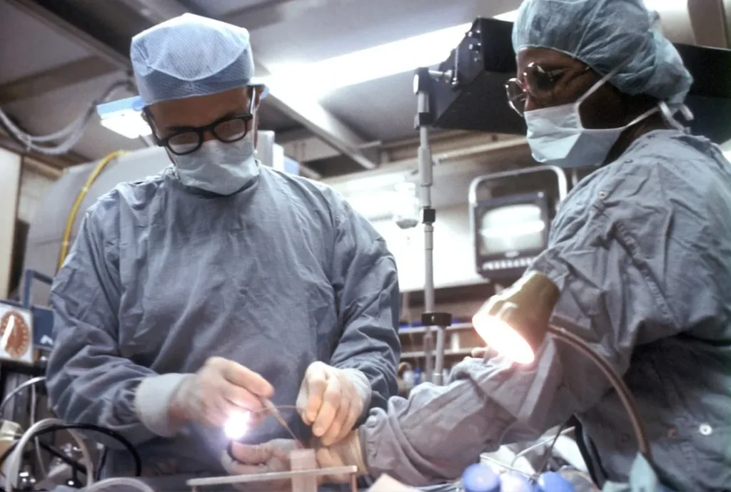
Spinal microsurgery frequently requires the use of long-shaft instruments such as suction tubes, Kerrison rongeurs, and nerve dissectors. The ANIM-Q Series features an extra-long working distance design, positioning the microscope further from the surgical site. This preserves high-definition image quality while providing surgeons with more generous instrument operating space. This design minimizes obstruction of the surgical field by the microscope body and enhances maneuverability in deep spinal canal and intervertebral foramen regions, particularly beneficial in complex decompression and fusion procedures.
Clinical Value:
- Liberates operating space in the deep surgical field
- Increases flexibility when using long instruments
- Reduces the risk of lens contamination
- Supports complex deep microsurgical manipulations
Flexible 6-Axis Electromagnetic Lock Stand
The ANIM-Q Series is equipped with a 6-axis electromagnetic lock stand system, enabling precise multi-directional positioning and rapid adjustments.
Clinical Value:
Polarized 4K 3D Medical Display System
The ANIM-Q Series includes a 55-inch polarized 4K 3D medical monitor as standard, providing the surgeon with a high-definition, ultra-low-latency, and true three-dimensional digital view. The pure exoscopic observation mode allows surgeons to operate without continuously looking through traditional eyepieces, promoting a more natural sitting or standing posture and improving ergonomic comfort. Simultaneously, assistants, scrub nurses, and teaching staff can share the 3D surgical field in real-time, enhancing team collaboration.
Clinical Value:
Improves surgeon posture and ergonomics
Enhances team collaboration efficiency
Supports digital education and training
Enables remote teaching and academic exchange
Stereoscopic Coaxial LED Cold Light Source
The ANIM-Q Series utilizes a high-brightness stereoscopic coaxial LED cold light source, providing uniform and stable illumination for the deep spinal surgical field. The cold light source effectively minimizes heat output, reducing the risk of thermal tissue damage, while improving the visualization quality of deep anatomical structures.
Provides uniform and stable illumination
Ensures true color reproduction
Enhances visualization of deep tissues

Suitable for prolonged microsurgical procedures
Common Spinal Conditions
Modern spinal surgery primarily addresses various degenerative and structural conditions using microsurgical techniques, with lumbar degenerative diseases being the most prevalent.
Lumbar Disc Herniation
Disc herniation compressing nerve roots can cause lumbago, leg pain, lower limb numbness, and muscle weakness. For patients unresponsive to conservative treatment or with significant nerve compression, microdiscectomy can precisely relieve neural compression and alleviate symptoms.
Lumbar Spondylolisthesis
Spondylolisthesis involves the displacement of one vertebra relative to another, leading to spinal stenosis and nerve compression. Patients may experience persistent low back pain, radiating leg pain, and limited mobility. As the condition progresses, spinal stability can be compromised, often necessitating decompression combined with interbody fusion and fixation.
Lumbar Spinal Stenosis
Lumbar spinal stenosis can compress the cauda equina or nerve roots, causing intermittent claudication, leg pain, and sensory disturbances. Microsurgical decompression techniques enable precise removal of compressive tissue while preserving spinal stability, restoring neurological function.
Cervical Spondylosis
Cervical degeneration can lead to compression of the spinal cord or nerve roots, resulting in upper limb numbness, pain, and motor dysfunction. Microsurgical cervical decompression and fusion procedures help relieve compression, restore neural conduction, and improve the patient's quality of life.
Surgical Microscope Empowering Precision Spinal Surgery
Digital surgical visualization is driving the evolution of spinal surgery from traditional optical microscopes towards intelligent, digital platforms. The ANIM Surgical Microscope, centered around Dual-Channel 4K 3D Imaging, Extra-Long Working Distance, Flexible 6-Axis Electromagnetic Lock Stand, Polarized 3D Display System, and Stereoscopic Coaxial LED Cold Light Source, constitutes a comprehensive digital solution tailored to the real-world clinical needs of spinal microsurgery. By delivering high-quality three-dimensional visualization, ample operating space, ergonomic exoscopic viewing, and team-shared visibility, it assists surgeons in more accurately identifying deep anatomical structures, enhancing operational stability and neural protection during complex spinal procedures. Furthermore, the system supports intraoperative high-definition image capture, case management, remote teaching, and digital operating room integration, providing ongoing support for clinical practice, education, and academic exchange. Addressing the demands of modern precision spinal surgery, Zhitong Technology is committed to leveraging digital surgical microscope technology to create a next-generation surgical vision platform for medical institutions, integrating precise visualization, meticulous operation, team collaboration, and digital management. This endeavor aims to guide complex spinal surgery towards a safer, more efficient, and intelligent future.