

Visualization is the foundation of precision in spine surgery.
Millimeter-level accuracy in a narrow corridor bordered by neural and vascular structures. The AINNOVI 3D exoscope platform is designed for the anatomical complexity of cervical, thoracic, and lumbar procedures.
The spinal canal demands more than traditional optics can deliver
The spinal canal is a narrow corridor. Every millimeter matters. Nerve roots, the dural sac, and segmental vessels sit within millimeters of the operative field.
Traditional operating microscopes have served the field for decades, but their bulk positioned directly over the surgical site restricts instrument maneuverability and forces the surgeon into a physically demanding posture. In minimally invasive approaches, the microscope competes with tubular retractor systems for the same physical space.
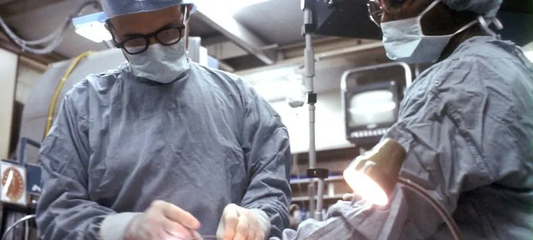
The exoscope separates the optical system from the surgeon. Camera positioned externally. Surgeon views the field on large monitors in a neutral, heads-up posture. The entire team sees the same 3D image.
70.4%
Of spine surgeons report musculoskeletal disorders from operative posture
100%
Of surgical teams report improved communication with shared 3D visualization

Three capabilities that change how spine surgery is performed
The exoscope is not a replacement for the operating microscope. It is an alternative visualization architecture with distinct advantages for specific procedural demands.
3D Microscopic Visualization
Passive polarization 3D technology delivers stereoscopic depth perception on large surgical monitors. The entire operating team — not just the primary surgeon — sees neural structures, vascular anatomy, and dural planes in true binocular depth.
- Neural identification: Nerve roots and dorsal root ganglia visualized with depth cues for safe dissection.
- Vascular assessment: Segmental arteries and epidural venous plexus perceived in three dimensions.
- Team synchronization: Assistant surgeon views the identical 3D field, enabling precise instrument coordination.
73.3%
Of surgeons rated 3D depth perception as excellent in a dual-center series of 880 spinal procedures.
Extended Working Distance
The camera is positioned extracorporeally, outside the surgical field and sterile draping. This creates an unobstructed volume for instrument insertion, manipulation, and exchange — particularly critical in minimally invasive tubular approaches where microscope bulk physically competes with retractor systems.
- Tubular retractor compatibility: Unrestricted maneuverability of screw drivers and cage inserters in MIS TLIF.
- Steep angulation access: Cervical corpectomy and lateral mass fixation without optical obstruction.
- Robotic positioning: Motorized arm with autofocus maintains optimal camera position during instrument exchanges.
100%
Of assistant surgeons reported enhanced awareness of the primary surgeon's maneuvers in published studies.
Integrated Fluorescence & Imaging
Native ICG video-angiography and 5-ALA fluorescence modules are integrated into the optical path — not added as external accessories. Real-time vascular and tumor boundary assessment without system switching or workflow interruption during intradural and complex spinal procedures.
- ICG angiography: Real-time arterial inflow and venous outflow assessment during DAVF ligation.
- 5-ALA tumor fluorescence: Tumor boundaries delineated from normal neural tissue in meningioma and schwannoma resection.
- Native 4K 3D recording: Fluorescence images recorded in the same format as white-light views for teaching and publication.
Zero
System switching required. ICG and 5-ALA filters engaged through software control without optical path interruption.
Procedures where visualization determines outcomes
Clinically validated across open and minimally invasive spinal approaches, from degenerative disease to intradural pathology.

Cervical Spine
- ACDF
- Cervical corpectomy
- Posterior laminectomy and fusion
- Lateral mass screw fixation
- OPLL resection
Thoracic Spine
- Lateral interbody fusion
- Decompression for disc herniation
- Burst fracture reduction
- Tumor resection
- Costotransversectomy
Lumbar Spine
- TLIF / PLIF / ALIF
- Bilateral decompression, unilateral approach
- Contralateral foraminotomy
- Revision decompression
- Interbody cage placement
Intradural & Complex
- Meningioma resection
- Schwannoma excision
- DAVF ligation
- Spinal cord tumor dissection
- Fluorescence-guided surgery
Operating microscope vs. 3D exoscope
Two architectures. Different strengths. The choice depends on procedural demands, surgeon preference, and OR workflow.
| Parameter | Operating Microscope | 3D Exoscope |
|---|---|---|
| Working Distance | Limited
Optics positioned directly over the surgical field. Bulk competes with retractor systems and instrument access in deep corridors. |
Extended
Camera extracorporeal. Unobstructed instrument maneuverability for tubular retractors, screw drivers, and cage inserters. |
| Surgeon Posture | Constrained
Neck flexion and shoulder abduction required to align with eyepieces. Documented risk factor for musculoskeletal disorders. |
Neutral
Heads-up posture facing monitors at eye level. 93.3% of surgeons report reduced fatigue in published studies. |
| Team Visualization | Restricted
Only the primary surgeon views the magnified field through binocular eyepieces. Assistant relies on indirect cues. |
Shared
Entire team views the same 3D image on large monitors. 100% report improved communication and assistant awareness. |
| Depth Perception | Monocular for team
Primary surgeon has stereoscopic view. Team views 2D on auxiliary monitor. Loss of depth for assistants and trainees. |
Stereoscopic for all
Passive polarization 3D on dual monitors. Full team perceives depth. Resident learning accelerated through shared perspective. |
| Fluorescence | Modular add-on
ICG and 5-ALA modules available as accessories. System switching may interrupt workflow during intradural procedures. |
Integrated
Native ICG and 5-ALA filter modules. Real-time vascular and tumor boundary assessment without system switching. |
| Learning Curve | Established
Decades of training infrastructure. Familiar to most spine surgeons. No additional technology adoption required. |
3 cases
Competency reported within 3 cases in clinical literature. 73.3% rated visualization as excellent from the first use. |
Clinical equivalence established: A 2026 meta-analysis of 785 patients across 7 comparative studies found no significant difference in operative time, complication rates, length of stay, or postoperative pain between 3D exoscopes and operating microscopes. Blood loss reduced by approximately 6 mL — statistically significant but clinically negligible. Source: Benato et al., 2026. PROSPERO CRD420251156505.
What the literature reports
Published data without selective emphasis. Evidence supports clinical equivalence with specific advantages in ergonomics and team workflow.

Meta-Analysis (2026)
No significant difference in operative time, LOS, complications, or postoperative pain. Blood loss reduced by ~6 mL.
Multicenter Series (2022-2025)
73.3% excellent visualization. Competency in 3 cases. 93.3% reduced fatigue. No conversions by 80%.
Systematic Review (2023)
All patients benefited clinically. Lumbar 48.6%. Discectomies 56.8%. Complications 2.6%. Switch rate 5.8%.
Limitations acknowledged: Current evidence consists primarily of retrospective and observational studies with small sample sizes and heterogeneous pathologies. The 2026 meta-analysis authors explicitly note that "larger, prospective trials are warranted." AINNOVI supports independent clinical research and welcomes collaborative study proposals.
Designed for the modern operating room
Compatible with standard OR infrastructure without proprietary lock-in.
Navigation Compatibility
Optical and electromagnetic navigation systems operate without interference. Camera positioning does not obstruct tracking arrays.
Universal Interface
Intraoperative Imaging
Robotic arm retracts automatically during fluoroscopy and C-arm acquisition. No manual repositioning required.
Auto-RetractNeurophysiological Monitoring
Video output compatible with standard neurophysiology displays. Independent monitor synchronized with primary display.
Multi-OutputDocumentation & Teaching
Native 4K 3D recording with surgeon's viewpoint. Direct export to PACS and education platforms. No external capture hardware.
Native RecordingExplore the platform
The AINNOVI visualization ecosystem for spine and beyond.
Q-Series
3D 4K external visualization platform with integrated fluorescence, robotic positioning, and extended working distance for spinal microsurgery.
View ProductEQ-Series
Ophthalmic surgical microscope with integrated OCT, digital imaging, and apochromatic optics for anterior and posterior segment surgery.
View ProductFind your support path
Request a surgical evaluation or download the clinical evidence summary for your department review.