

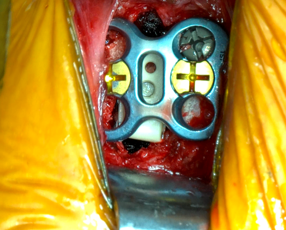
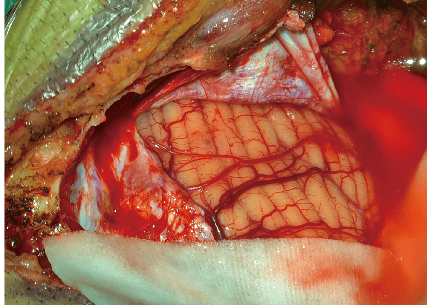
Spine surgery has always been one of the most demanding specialties in modern surgery, requiring exceptional precision. With the advancement of minimally invasive spine surgery, complex spinal reconstruction, and spinal tumor surgery, surgeons must perform highly delicate tissue dissection, neural protection, and precise implant placement within extremely confined operating spaces.
Traditional optical surgical microscopes have long been an essential visualization tool in spine surgery. However, as surgical complexity continues to increase, conventional microscopes are increasingly showing their limitations:
Surgeons must maintain a prolonged head down posture through eyepieces, leading to neck and shoulder fatigue;
The field of view is constrained by the microscope’s mechanical structure;
The surgical team cannot share the same high definition visual feed;
Depth perception in deep, narrow spaces still leaves room for improvement.
In recent years, 3D exoscopes based on digital imaging technology have been driving a paradigm shift in spine surgery visualization platforms, offering new solutions for the next generation of precision surgery.
1.What Is 3D Exoscopic Technology?
3D exoscopic technology is an innovative surgical visualization platform built on high definition digital camera systems, three dimensional stereoscopic imaging, and large screen display technology. Unlike traditional microscopes that rely on direct observation through eyepieces, a 3D exoscope captures images of the surgical field via a high resolution camera mounted on a robotic arm, then processes and displays real time 3D images on a 3D high definition monitor.
Its core technologies include:
High Resolution Digital Imaging
Modern 3D exoscopes employ high magnification, high sensitivity imaging technology to deliver clear tissue layer delineation, enabling surgeons to visualize:
Nerve root structures;
Dural sac boundaries;
Intervertebral disc tissues;
Bony anatomical landmarks;
Small blood vessels and soft tissues.
Stereoscopic 3D Vision
In spinal surgery, depth perception is a critical factor affecting procedural safety. 3D visualization systems use dual channel imaging to simulate human binocular vision, providing surgeons with true spatial awareness — more accurate judgment of tissue distances, enhanced identification of deep structures, and support for fine dissection and precise manipulation. This enhanced spatial perception is particularly valuable when working near high risk structures such as the spinal cord and nerve roots.
2.How Does 3D Visualization Improve Precision in Spine Surgery?
Enhanced Identification of Complex Anatomical Structures
The spinal region has highly intricate three dimensional anatomy. For example:
The cervical spine is adjacent to the spinal cord and vertebral arteries;
The thoracic spine offers narrow working spaces;
The lumbar spine involves nerve root protection and interbody fusion procedures.
Traditional 2D visualization tends to compromise depth judgment. 3D exoscopic technology, by providing stereoscopic imaging, enhances spatial relationships among tissues, allowing surgeons to intuitively comprehend complex anatomy.
Studies have shown that 3D visualization techniques can improve surgeons’ ability to judge depth and spatial relationships, and enhance visual feedback quality during complex minimally invasive procedures.
Supporting the Development of Minimally Invasive Spine Surgery
The trend in minimally invasive spine surgery is toward: smaller incisions, less tissue trauma, and greater procedural precision. However, smaller incisions mean: reduced operating space, limited visual fields, and more challenging instrument maneuverability.
3D exoscopes, through digital magnification, high magnification display, and flexible camera positioning, provide surgeons with greater visual freedom. Particularly in:
Percutaneous pedicle screw placement;
Endoscopic assisted foraminotomy;
Lumbar decompression;
Spinal fusion procedures,
high definition 3D vision enables surgeons to perform critical steps with greater accuracy.
3.How Does Extended Working Distance Transform Spine Surgery?
What Is Working Distance?
Working distance is the distance between the microscope’s optical system and the surgical target area. For spine surgery, a greater working distance means:
More generous instrument maneuvering space;
Greater flexibility in surgical angles;
Fewer instrument collisions;
Better access to deep regions.
Advantages of Extended Working Distance in Spine Surgery
Enhanced Freedom in Deep Spinal Regions
Spine surgery often requires operating in deep, narrow spaces, such as:
Spinal canal decompression;
Nerve root release;
Dural protection;
Tumor resection.
Traditional microscopes, with their relatively short working distance, can restrict instrument movement due to the proximity of the lens to the surgical field. 3D exoscopic systems featuring an extended working distance of over one meter allow the camera to be positioned farther away while maintaining high magnification clarity. This affords surgeons significantly greater working room.
Improved Surgeon Ergonomics
When using traditional microscopes for extended periods, surgeons must maintain a fixed posture through the eyepieces. Prolonged procedures can lead to:
Neck strain;
Shoulder fatigue;
Lower back discomfort.
3D exoscopic systems adopt a screen based viewing mode: surgeons can maintain a more natural, upright posture while operating via a large format 3D display. This ergonomic advantage is especially valuable during lengthy spinal surgeries.
Facilitating Team Collaboration and Education
Traditional microscopes are primarily designed for a single surgeon. Digital 3D exoscopic platforms, on the other hand, can:
Allow assistants to simultaneously view the surgical field;
Support teaching and training;
Enable remote consultation;
Record high definition surgical footage.
Looking ahead, with the development of artificial intelligence (AI), augmented reality (AR), and smart operating rooms, digital visualization platforms will become a vital component of the intelligent surgical suite.
4.Future Trends: 3D Visualization as a Core Visualization Platform in Spine Surgery
As digital healthcare technology advances, surgical microscopes are evolving from traditional optical devices toward digital, intelligent systems. The next generation spine surgery visualization platform will not only provide magnified images but will also integrate:
AI assisted anatomical identification;
Surgical navigation;
Real time data fusion;
Telemedicine;
Intelligent teaching systems.
3D exoscopic technology, as a key direction in digital surgical microscopy, is poised to become essential infrastructure for future precision spine surgery. For medical device companies, developing clearer, smarter, and more ergonomic visualization solutions will further enhance surgeons’ capabilities in complex operative environments.
Conclusion
3D visualization technology is redefining the visual paradigm of spine surgery. By delivering:
High resolution 3D imaging;
Enhanced spatial depth perception;
Extended working distance design;
Optimized ergonomics;
Digital team collaboration capabilities,
it enables spine surgeons to achieve more precise, safer, and more efficient surgical outcomes.
As digital operating rooms continue to evolve, 3D visualization technology will become a cornerstone of next generation spine surgery visualization platforms.