

Otology is one of the most visually demanding specialties in modern surgery.With the advancement of minimally invasive techniques, otological procedures are progressively shifting from traditional open approaches toward more precise, less invasive, and function preserving strategies. Commonly performed ENT surgeries include:
Middle and inner ear microsurgery;
Skull base tumor resection;
Acoustic neuroma excision;
Facial nerve decompression.
These procedures share several common characteristics:
Extremely confined operating spaces;
Highly complex anatomical structures;
Dense concentrations of critical nerves and vessels;
Surgical pathways involving numerous challenging angles.
As a result, surgeons require not only high definition visualization but also true three dimensional spatial awareness.
Conventional optical microscopes have long supported the development of ENT microsurgery. However, as the demands of complex otological procedures continue to grow, digital 3D exoscopes are emerging as a key direction for next generation surgical visualization platforms.
1.What Is a 3D Exoscope?
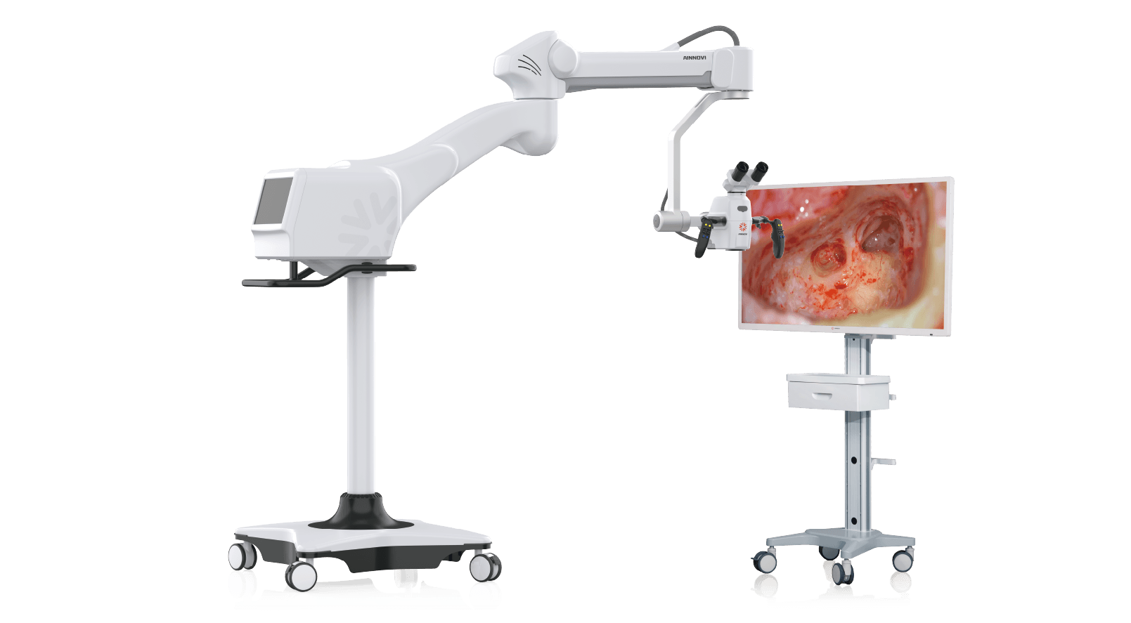
A 3D exoscope is an innovative surgical visualization system that integrates:
High definition digital camera technology;
Three dimensional stereoscopic imaging;
Digital image processing;
Large screen 3D display technology.
Unlike traditional microscopes that require direct observation through eyepieces, a 3D exoscope captures images via an ultra HD camera system positioned above the surgical field and displays real time 3D imagery on a high definition 3D monitor.
This paradigm shifts the way surgeons view the operative field — transforming visualization from “single surgeon observation” to “entire surgical team sharing ultra HD 3D visual information.”
2.The New Challenges in Visual Technology for Otology
Conventional optical microscopes have long been essential tools in otological microsurgery, providing surgeons with natural 3D stereoscopic vision through binocular optical systems. However, as otological procedures evolve toward greater precision, the application model of traditional microscopes faces new challenges.
First, in complex ear surgeries, surgeons must not only observe magnified local structures but also integrate spatial relationships, instrument trajectories, and deep zone anatomy into their overall assessment. Second, because observation is conducted through eyepieces, the visual field is largely limited to the primary surgeon — assistants, nurses, and trainees cannot simultaneously access equally clear stereoscopic images, which hampers team collaboration and surgical education. Third, prolonged fine manipulation through eyepieces requires surgeons to maintain a fixed head and neck posture, leading to cervical and shoulder fatigue that compromises long term surgical comfort.
Traditional optical microscopes rely on the surgeon’s experience based integration of visual information through eyepiece observation. In contrast, digital visualization systems can further combine high definition imaging, image processing, and large screen display technologies to deliver a more open, shared, and intelligent visual experience. 3D exoscopes, by capturing surgical field data through ultra HD dual channel camera systems and presenting real time stereoscopic images on 3D displays, enable the entire surgical team to share consistent stereoscopic information, offering a novel visualization solution for otological microsurgery.
3.How Does a 3D Exoscope Enhance Precision in Otological Procedures?
Enhanced Spatial Awareness and Depth Perception
In microsurgery, accurate spatial perception directly impacts both procedural safety and surgical outcomes. While conventional optical microscopes provide stereoscopic vision, surgeons still rely heavily on eyepiece based observation for spatial judgment. In complex, confined otorhinolaryngological and skull base regions — characterized by deep anatomical structures, delicate tissue layers, and micro instrument manipulation — surgeons must still combine experience with spatial orientation and distance estimation.
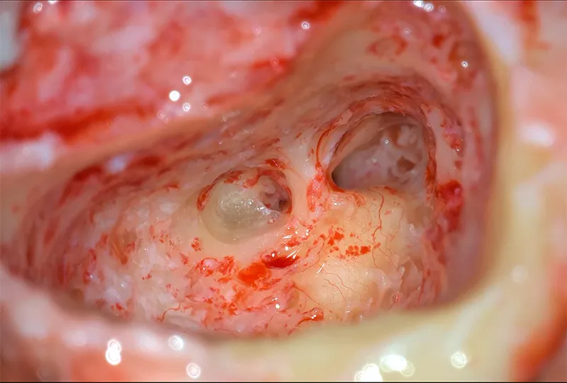
3D exoscopes, through dual channel 4K3D imaging technology and digital image processing, generate ultra HD 3D stereoscopic images. Surgeons can now intuitively perceive spatial distances between tissues, positional relationships between instruments and critical anatomical structures, and layered relationships in deep regions on a large format 3D display.
This enhanced spatial feedback helps improve intraoperative precision and reduce procedural risks in complex anatomical zones — particularly valuable in otology, where exacting spatial accuracy is paramount.
Multi Directional Observation at Challenging Angles
One of the greatest challenges in otological surgery is that pathological targets are often concealed within complex anatomical spaces. Many lesions are located deep, surrounded by vital structures, and accessible only through limited working corridors. To achieve optimal viewing angles, surgeons must frequently adjust microscope position, tilt, or patient positioning — adding complexity to the procedure — while being constrained by the optical path and eyepiece orientation.
3D exoscopes, leveraging dual channel 4K3D imaging combined with a flexible, user friendly stand system, allow surgeons to observe the surgical field on a large format 3D display while maintaining a more natural and comfortable posture. Camera angles can be rapidly adjusted as needed to view the target area from different directions.
Compared to conventional microscopes that rely on optical path adjustments, 3D exoscopes significantly enhance field of view flexibility, deep structure visualization, and instrument maneuverability in complex anatomical regions, delivering more open and digitally enabled visual support for high precision ear surgery.
Enabling Precise Anatomical Preservation
A critical objective in ENT surgery is not only lesion removal but also preservation of patient function. For example:
Skull base surgery requires protection of the optic nerve, internal carotid artery, cranial nerves, and dural structures;
Otological surgery demands preservation of the facial nerve, auditory structures, and vestibular function.
3D high definition visualization helps surgeons more clearly identify normal tissue boundaries, pathological regions, and neurovascular relationships, thereby supporting more precise and safer surgical execution.
4.Otology 3D Exoscopes: Driving the Digital OR Transformation
Digital microscopes are changing not only how surgeons visualize but also how surgical teams collaborate.
Improved Surgeon Ergonomics
Traditional microscopes require surgeons to remain close to the eyepieces for extended periods, maintaining fixed head and neck postures with sustained visual concentration. Prolonged complex procedures can lead to neck and shoulder fatigue and increased physical strain.
3D exoscopes, by enabling observation through 4K ultra HD displays, allow surgeons to operate in a natural seated posture with a more open visual environment, thereby improving ergonomic performance during lengthy procedures.
Enhanced Teaching and Team Collaboration
Conventional microscopes primarily serve the lead surgeon, limiting the viewing range and teaching efficiency. Digital 3D systems, in contrast, enable assistant surgeons, residents, medical students, and nursing teams to simultaneously view high definition 3D imagery — a significant advantage for surgical training, academic exchange, and remote mentoring.
5.Conclusion: 3D Exoscopes Driving Precision in Otology
As otological surgery continues its trajectory toward minimally invasive and precision focused approaches, visualization technology is becoming a decisive factor in surgical quality. 3D exoscopes empower ENT surgeons to achieve more precise, safer, and more efficient procedures in challenging angles and confined spaces.
Looking ahead, as digital operating rooms continue to evolve, 3D exoscopes will become an integral component of the otolaryngology surgical visualization landscape.